X-RAY Photoconductivity Setup

X-ray Photoconductivity Setup

Daniele M. Trucchi  -

DiaTHEMA Lab

 
The x-ray photo-detection setup is designed to allow measurements under continuous (DC) and modulated radiation (AC) exposition. In this way it is possible to derive the current flowing into the devices, the total current under irradiation and the net photo-generated current, at the variation of modulation frequency, bias voltage and radiation dose-rate. The x-ray beam can be filtered by a primary and/or a variable thickness secondary filter. The beam impinges orthogonally on the device. The detector is biased by a voltage generator: in case of continuous excitation, the dark and photo-generated current are measured by a pico-ammeter, while in modulated excitation, the net current signal is amplified and converted in a voltage signal by a current-voltage converter and sent to a lock-in amplifier, which compares the signal of interest with the reference one generated by the chopper controller. The modulated electronic chain implies, with respect to continuous irradiation conditions, the advantage of: High SNR discrimination of signal, Independence of device leakage current for a broad current range (up to 0.1 mA). This means that, under this operative condition, we can both evaluate the devices behaviour in DC irradiation (small modulation frequencies) and devices performance for AC applications. All the setup is controlled by a computer via GPIB and RS-232 buses which has the function of collecting data and controlling the electronic instruments. A ionization chamber designed and calibrated for photons < 100 keV was recently introduced within the setup. The aim is to continuously monitor the source intensity, to compare the device under test performance with the dosimetric benchmark detector and to perform absolute dose measurements.


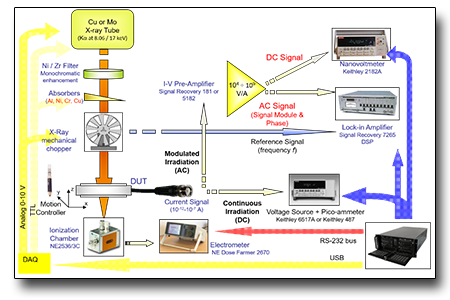
TECHNICAL SPECIFICATIONS

  • Mo or Cu tubes;

  • Accelerating voltage up to 60 kV, current up to 60 mA, max 3 kW;

  • Continuous wave or modulated operations;

  • modulation frequency up to 800 Hz;

  • microfocused beam for mapping with 0.1 mm resolution;

  • 3-axes + rotation stages

  • Completely automated operations
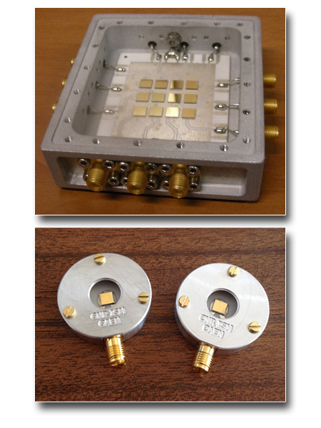


SAMPLES

  • maximum size: 50x50mm2
 

USE FOR

  • wide bandgap semiconductors;
  • perovskites;
  • detectors;
  • dosimeters;
  • photovoltaic cells


CASE STUDIES

Development of diamond dosimeters for ionizing radiation and neutron detectors.

The activity of developing and characterizing the response of radiation-hard, sensitive, and tissue equivalent detectors made of synthetic diamond is a plurennial activity of DiaTHEMA Lab.

The main reference manuscripts are:

D.M. Trucchi, P. Allegrini, P. Calvani, A. Galbiati, K. Oliver, and G. Conte, Very Fast and Priming-less Single-Crystal Diamond X-ray Dosimeters, IEEE Electron Device Letters, 33 (2012) 615-617.

A. Bellucci, S. Orlando, D. Caputo, E. Cappelli, and D.M. Trucchi, Dosimetric Performance of Single-Crystal Diamond X-ray Schottky Photodiodes, IEEE Electron Device Letters 35 (2013) 695-697

M. Girolami, A. Bellucci, P. Calvani, C. Cazzaniga, M. Rebai, D. Rigamonti, M. Tardocchi, M. Pillon and D.M. Trucchi, Mosaic diamond detectors for fast neutrons and large ionizing radiation fields, Physica Status Solidi A 212 (2015) 2424–2430.


We use cookies essential for the functioning of the site. You can decide for yourself whether or not to allow cookies. Please note that if you refuse them, you may not be able to use all of the site's features.