Research Activities
The group’s research activity has focused on the characterization of nanostuctures and nanosystems with interest in biology and medicine, through scanning probe microscopies and high resolution techniques. The group has also performed studies of structure and function of biosystems employing different spectroscopy techniques, such as Raman, vis/IR, XANES and X-ray.
Regarding the SPM studies, we have evaluated the alterations induced by stress stimuli on biological systems (pathological, pharmacological, and pollution).
The group has often explored innovative avenues to improve the understanding of the biological world. For instance, at Biotech@ISM we have developed a novel nanomechanical sensor, the nanomotion sensor, to characterize in real-time the metabolic activity of biological specimens.
Projects
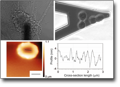
Development of a Nanomotion sensor to study cells and bacterial metabolic activity.
The BioTech@ISM group of the Istituto di Struttura della Materia - CNR in Rome (ISM-CNR) has developed a nanomotion sensor based on nanomechanical oscillators, which is able of measuring the nanoscale vibrations of single cells in physiological conditions (Mustazzolu et al., Antimicrobial Agents and Chemotherapy, 2019). This instrument is based on a commercial Atomic Force Microscope (AFM), mounted on an inverted optical microscope and connected to an I/O card for direct collection of the metabolically-induced cellular movement into a measurable nanomotion signal. In most cases, the sensor is a commercial AFM probe, while the nanomotion signal is obtained by modifying the instrument to allow monitoring of the sensor deflection. Such signal provides real-time, correlated information on the metabolic activities of the cells that occur in response to physical, biological or chemical stimuli directly deliverable in the measurement chamber.
The methodology has been validated in studies on bacteria and mammalian cells, and was exploited in applications that range from microbiology to oncology and astrobiology (Venturelli et al., J. Mol. Recog., 2020). A recent study focused on the interaction of beta-amyloids with neurons and highlighted an important role of the nanomotion sensor in the field of neurodegeneration (Vannocci et al., Sci. Rep., 2019).
A perspective view on the nanomotion detection of living organisms and its features
Journal of Molecular Recognition, e2849 (2020)
A new tool to determine the cellular metabolic landscape: nanotechnology to the study of Friedreich’s ataxia
SCI REP. 9, 19282 (2019)
A rapid unravelling of mycobacterial activity and of their susceptibility to antibiotics
ANTIMICROBIAL AGENTS AND CHEMOTHERAPY, vol. 3, p. 2194 (2019)
Human erythrocyte's ageing in healthy and pathological subjects, studied by Differential Scanning Calorimetry, Atomic Force Microscopy and biochemical patterns.
In this Project, two research groups from the institutions have put together their expertise to study the ageing process of RBC from healthy and pathological subjects. To this end we have combined the results collected through specific microscopy and spectroscopy techniques, AFM and DSC, with the evaluation of several biochemical parameters.
The idea is to identify and characterize, by DSC measurements, the main proteins involved in the ageing process at three different levels of complexity: whole cells, intracellular content and isolated membrane-skeleton. This information has been coupled with the AFM quantitative measurements of morphometric and morphomechanic parameters of the whole cells and of the isolated membranes.
The goal of these characterizations is to carry out a pattern typical of the ageing path in healthy and pathologic condition and whose differences can be used to gain information of clinical relevance.
Identification of Oxidative Stress in Red Blood Cells with Nanoscale Chemical Resolution by Infrared Nanospectroscopy.
INT. J. MOL. SCI., 19, 2582 (2018).
Insights into the morphological pattern of erythrocytes' aging: Coupling quantitative AFM data to microcalorimetry and Raman spectroscopy.
JOURNAL OF MOLECULAR RECOGNITION, vol. 31 (11), e2732.
Nanosized metal pollutant in mytilus tissues.
This study was part of a marine monitoring program developed by ENI s.p.a. in order to control the environmental effects associated to the building of gas extraction platforms in the Adriatic Sea.
Concerning the microscopy, the study required morphological, rheological and nanomechanical AFM analysis of histological sections of tissues extracted from the most sensitive target organs of the mytili. During the study, the presence of nanomaterials was evidenced, mostly, by lateral force images while the collection of force curve maps highlighted the ability of some pollutants to induce local alterations in the nano-mechanical properties of the tissues. The degree of local alteration of the organs’ texture was also assessed.
Metal‐based micro and nanosized pollutant in marine organisms: What can we learn from a combined atomic force microscopy‐scanning electron microscopy study.
Journal of Molecular Recognition (2020)
Instrumentation
Collaborations
- King’s College (London, UK) Prof. A. Pastore; Dr. T. Vannocci
- EPFL (Lausanne, CH): Prof. G. Dietler; dr S. Kasas
- Bulgarian Academy of Science (Dip. Biophysics and Biomedical engineering; Prof. S. Taneva; dr. S. Krumova)
- CNR: IFT (Roma, Dr. A. Lisi); IRBIM (Ancona, Dr. G. Fabi)
- University Tor Vergata (Dip. Biology) Prof. S. Cannata
- University of Cassino (Prof. F. Misiti)
- University of Rome, La Sapienza (Dip. Biochemical Sciences; Prof. A. Bellelli; Dr. G. Boumis)
- Delft University, Delft, NL (Prof. Farbod Alijani)
 Italiano (Italia)
  English (UK)