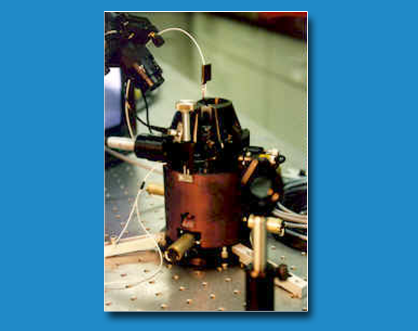
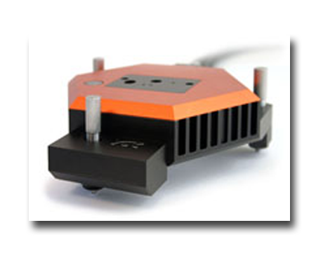
Scanning probe microscope sNanosurf mod. FlexAFM and AFM home designed
Marco Girasole -
Giovanni Longo -
BioTech@ISMb
Several instruments are, or can be, coupled to the microscopes to allow a more complete analysis of the samples. In particular, the AFM is mounted on an inverted optical microscope potentially enabling fluorescence analysis and can be coupled to external, even femto-sec, laser sources. Concerning the study of biosystems, or other samples that require environmental control, the microscopes can be integrated in an incubator which guarantees controlled and ideal conditions for cell growth.
TECHNICAL SPECIFICATIONS
- Fast flexural scanning stage
- Open or closed loop X/Y/Z feedback
- Probe monitoring through IR laser (800nm)
- Max scanning area (x/y/z): 100x100x20 µm
- Max sample size (x/y/z): fino a 5x5x0.5 cm
- Max Force Curve collection speed: 0.05 s
- Max imaging scanning speed: 0.5 sec/row
AVAILABLE TECHNIQUES
- Morphological properties in contact mode, non-contact mode, tapping mode
- Lateral force imaging of the samples (i.e. rheological prop.) with nanometer resolution
- Electric and magnetic force imaging
- Collection of single, multiple and maps of force-distance curves
- Nanomotion sensors
SAMPLE
- The AFM allows collecting morphological images and maps of nanomechanical properties with nanometer resolution in different environmental conditions, including liquids. No sample’s electrical properties are required.
Coupling the AFM characterization with appropriate spectroscopic investigations, it is possible to effectively characterize monolayers (functionalized surfaces, sensors/biosensors), two-dimensional systems, organic-inorganic systems of interest for energy harvesting, macromolecular complexes, folding/unfolding dynamics, protein alterations in solution or can be used to follow the temporal evolution of the samples
USE FOR
-
Imaging the samples morphology with nanometer resolution and in time laps
-
Imaging the tribological properties of the samples (lateral force imaging)
-
Imaging the electric and magnetic properties of the samples on the nanoscale
-
Measure the nanomechanical properties of the samples (stiffness, elasticity, Young’s modulus etc) on the nanoscale
-
Measure the viscoelastic behaviour of the samples
-
Measure of forces (adhesive, idration, double layer, hydrophobic, covalent bonding etc.)
-
Perform Force spectroscopy of molecule and polymers
-
Measure the nano-motility of biological systems (nanomotion sensors)
Case Studies
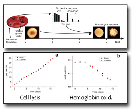
Erythrocytes aging in normal and microgravity conditions
Microgravity is, for highly perceptive cells such as RBCs, an environmental stimulus that is sensed and processed as well as chemical stimuli. In this work we studied, for the first time, the aging in microgravity for cells that live in suspension. The results showed that in microgravity the cell aging follows a different path compared to standard condition. Indeed, microgravity is sensed by the cell as a biomechanical alteration, then is translated into modulations of the metabolic cell cycles and finally results in distinct morphological patterns compared to the normal aging. The study required the combined use of AFM, biochemical and UV/vis techniques.
See: Dinarelli et al. Scientific Report 8:5277 (2018)
Nanosized metal pollutant in mytilus tissues
This study was part of a marine monitoring program developed by ENI s.p.a. in order to control the environmental effects associated to the building of gas extraction platforms in the Adriatic sea. Concerning the microscopy, the study required morphological, rheological and nanomechanical AFM analysis of histological sections of tissues extracted from the most sensitive target organs of the mytili. During the study, the presence of nanomaterials was evidenced, mostly, by lateral force images while the collection of force curve maps highlighted the ability of some pollutants to induce local alterations in the nano-mechanical properties of the tissues. The degree of local alteration of the organs’ texture was also assessed.
See: Dinarelli et al. J. Mol. Rec. e2851 (2020)
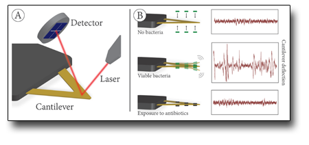
Nanomotion sensor: the motion of biosystems at the nanoscalea
Exploiting the AFM's ability to translate small vertical displacements of the cantilever into measurable signals, the AFM microscopes of BioTech@ISM have been modified to measure the nanometric vibrations of living biological systems. This is an innovative application of the AFM, the nanomotion sensor, which is proposed as a new real-time investigation tool of the behavior of biosystems and of their metabolic activity. The nanomotion sensor has a wide spectrum of applications ranging from the study of enzymatic cycles of proteins, to microbiology up to strategies for fighting against neurodegenerative diseases.
See: Dinarelli et al. J. Microbiol. Met. 138:72 (2017)
 Italiano (Italia)
  English (UK)