Antonio Cricenti -
Marco Luce -
Marco Ortenzi -
David Becerril - Borsista
Stefano Bellucci (INFN) - Associato ISM
Research Activities
The activity is divided into the studies of samples of materials and biological science, and clean semiconductor/metal surfaces and the development of prototype.
In details:
As for local probe scanning microscopy, all the instruments are STM-AFM-SNOM prototypes developed in the institute, that can observe the same area of the sample in different operating modes (contact, non-contact, friction or phase shift mode). In particular, the SNOM can use different optical set-ups (tip lighting, tip collection or lighting / collection modes) optimized to adapt to specific experimental needs.
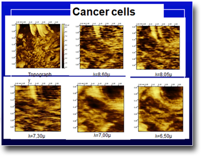
Reflectivity of cancer cells at various infrared wavelengths
Localized internal photoemission, reflectance, transmission, fluorescence can be performed in the UV-IR range (lambda 0.4-10 microns). Any type of sample can be observed in a range from a few nm to half a millimeter with chemical sensitivity as well as topographical.
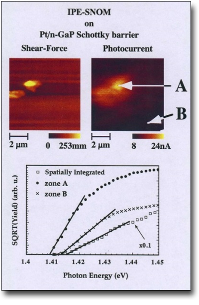
Measurement of the Schottky barrier in the Pt / n-GaP system by means of internal photoemission located in different points of the sample
Angular resolved photoelectron spectroscopy (ARUPS) is a surface-sensitive quantitative spectroscopic technique that measures the energy of the emitted electrons and their emission angle and, consequently, the information of the vector k. The spectra are obtained by irradiating a material with a beam of UV rays (20-40 eV) and simultaneously measuring the number of electrons with a certain kinetic energy which emerges from the last layers (0-10 nm) of the analyzed material.
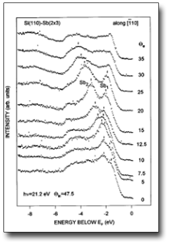
Example of photoemission spectra as a function of the emission angle for the surface Si (110) -Sb (2x3)
Auger and XPS techniques resolved angularly measure the elemental composition of a few parts per thousand of materials and are obtained by irradiating a material with an electron beam (Auger) or X-rays (XPS) and simultaneously measuring the kinetic energy and the number of electrons at any emission angle, in order to be more sensitive to the surface or bulk.
Relevant Publications
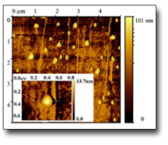 AFM image of stabilized gold nanoparticles
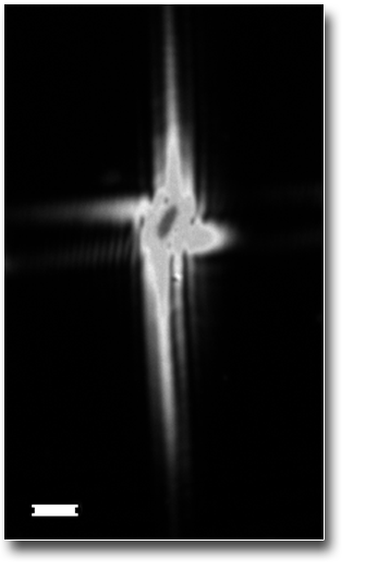 Fluorescence image of the FERMI-XFEL focused beam
Bonfigli, F.; Capotondi, F.; Cricenti, A., Giannessi, L.; Kiskinova, M.; Luce, M.; Mahne, N., Manfredda, M.; Montereali, R.; Nichelatti, E.; Pedersoli, E.; Raimondi, L.; Vincenti, M.A.; Zangrando, M.; (2019)
“Imaging detectors based on photoluminescence of radiation-induced defects in lithium fluoride for XFEL beam monitoring"
Il Nuovo Cimento C
“Imaging detectors based on photoluminescence of radiation-induced defects in lithium fluoride for XFEL beam monitoring"
Il Nuovo Cimento C
Callari, Giuseppina; Mencattini, Arianna; Casti, Paola, Comes, Maria Colomba; Di Giuseppe, Davide; Di Natale, Corrado; Sammarco, Innocenzo; Pietroiusti, Antonio; Magrini, Andrea; Lesci, Isidoro Giorgio; Luce, Marco; Cricenti, Antonio; Martinelli, Eugenio (2019)
“The Influence of Uncertainty Contributions on Deep Learning Architectures in Vision-Based Evaluation Systems in IEEE transactions on instrumentation and measurement"
“The Influence of Uncertainty Contributions on Deep Learning Architectures in Vision-Based Evaluation Systems in IEEE transactions on instrumentation and measurement"
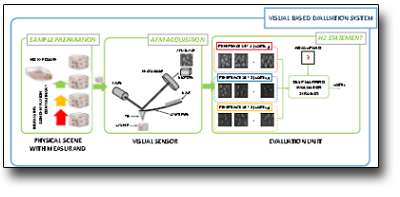 Digitization scheme of AFM images
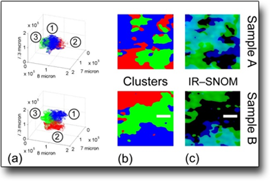 Study of cancer and control cells of the base of the esophagus using localized infrared reflectivity.
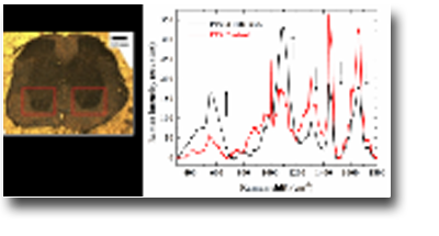 Spinal nerve microRaman analysis 75 days after infection.
 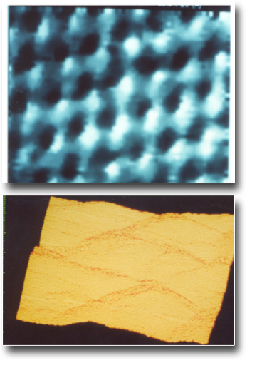 STM image of the graphite surface cleaved in air (left) and of a gold grating (right)
Instrumentation
AFM@Nanospectroscopy SNOM XPS@Nanospectroscopy ARUPS
Collaborations
- UNAM Messico,
- Fondazione Santa Lucia,
- Università di Liverpool,
- Dipartimento di Fisica, Ingegneria Elettronica e Biomedicina e Prevenzione dell’Università Roma Tor Vergata,
- INFN,
- ENEA,
- Dipartimento di Fisica Università di Roma La Sapienza e Roma3,
- Vinca Institute Belgrad.
 English (UK)
  Italiano (Italia)